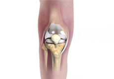
Knee Anatomy
The knee is a complex joint made up of different structures - bones, tendons, ligaments, and muscles. They all work together to maintain the knee’s normal function and provide stability to the knee during movement.
Having a well-functioning healthy knee is essential for our mobility and ability to participate in various activities. Understanding the anatomy of the knee enhances your ability to discuss and choose the right treatment procedure for knee problems with your doctor.
Bones of the Knee
The knee is a hinge joint made up of two bones, the thighbone (femur) and shinbone (tibia). There are two round knobs at the end of the femur called femoral condyles that articulate with the flat surface of the tibia called the tibial plateau. The tibial plateau on the inside of the leg is called the medial tibial plateau and on the outside of the leg, the lateral tibial plateau.
The two femoral condyles form a groove on the front (anterior) side of the knee called the patellofemoral groove. A small bone called the patella sits in this groove and forms the kneecap. It acts as a shield and protects the knee joint from direct trauma.
A fourth bone called the fibula is the other bone of the lower leg. This forms a small joint with the tibia. This joint has very little movement and is not considered a part of the main joint of the knee.
Articular Cartilage and Menisci of the Knee
Movement of the bones causes friction between the articulating surfaces. To reduce this friction, all articulating surfaces involved in the movement are covered with a white, shiny, slippery layer called articular cartilage. The articulating surface of the femoral condyles, tibial plateaus and the back of the patella are covered with this cartilage. The cartilage provides a smooth surface that facilitates easy movement.
To further reduce friction between the articulating surfaces of the bones, the knee joint is lined by a synovial membrane that produces a thick clear fluid called synovial fluid. This fluid lubricates and nourishes the cartilage and bones inside the joint capsule.
Within the knee joint, between the femur and tibia, are two C-shaped cartilaginous structures called menisci. Menisci function to provide stability to the knee by spreading the weight of the upper body across the whole surface of the tibial plateau. The menisci help in load-bearing i.e. it prevents the weight from concentrating onto a small area, which could damage the articular cartilage. The menisci also act as a cushion between the femur and tibia by absorbing the shock produced by activities such as walking, running and jumping.
Ligaments of the Knee
Ligaments are tough bands of tissue that connect one bone to another bone. The ligaments of the knee stabilize the knee joint. There are two important groups of ligaments that hold the bones of the knee joint together, collateral and cruciate ligaments.
Collateral ligaments are present on either side of the knee. They prevent the knee from moving too far during side to side motion. The collateral ligament on the inside is called the medial collateral ligament (MCL) and the collateral ligament on the outside is called the lateral collateral ligament (LCL).
Cruciate ligaments, present inside the knee joint, control the back-and-forth motion of the knee. The cruciate ligament in the front of the knee is called anterior cruciate ligament (ACL) and the cruciate ligament in the back of the knee is called posterior cruciate ligament (PCL).
Muscles of the Knee
There are two major muscles in the knee - the quadriceps and the hamstrings, which enable movement of the knee joint. The quadriceps muscles are located in front of the thigh. When the quadriceps muscles contract, the knee straightens. The hamstrings are located at the back of the thigh. When the hamstring muscles contract, the knee bends.
Tendons of the Knee
A tendon is a tissue that attaches a muscle to a bone. The quadriceps muscles of the knee meet just above the patella and attach to it through a tendon called the quadriceps tendon. The patella further attaches to the tibia through a tendon called the patella tendon. The quadriceps muscle, quadriceps tendon, and patellar tendon all work together to straighten the knee. Similarly, the hamstring muscles at the back of the leg are attached to the knee joint with the hamstring tendon.
Knee Conditions
Knee Fracture
A fracture is a condition in which there is a break in the continuity of the bone. In younger individuals, these fractures are caused by high energy injuries, as from a motor vehicle accident. In older people, the most common cause is a weak and fragile bone.
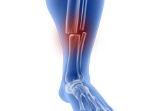
Fractures of the Tibia
The lower leg is made up of two long bones called the tibia and fibula that extend between the knee and ankle. The tibia or shinbone is the larger of the two bones. It bears most of the body’s weight and helps form the ankle joint and knee joint.
Fractures of the Patella
The patella or kneecap is a small bone present in the front of your knee where the thigh bone meets the shinbone. It provides protection to your knee and attachment to muscles in the front of the thigh. An injury to the knee can result in a break or fracture of the patella.
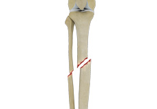
Tibial Shaft Fracture
The lower leg is made up of two long bones called the tibia and fibula that extend between the knee and ankle and help form the ankle joint and knee joint. The tibia or shinbone is the larger of the two bones and is one of the major bones of the lower leg. It bears most of the body’s weight and plays a crucial role in balancing body weight when standing and walking.
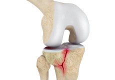
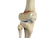
Tibial Plateau Fracture
A tibial plateau fracture is a crack or break on the top surface of the tibia or shinbone in the knee joint. The fracture most often occurs following a high-intensity trauma or injury from the impaction of the femoral condyles over the tibial plateau.
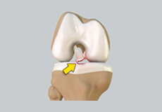
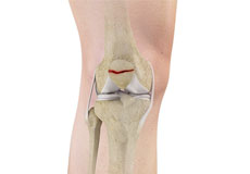
Tibial Eminence Fractures
The tibial eminence, also called the tibial spine, is a bony protuberance of the tibia (shin bone) that attaches to the anterior cruciate ligament (ACL) of the knee joint.
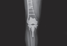
Periprosthetic Knee Fractures
Knee replacement, also called knee arthroplasty, is a surgical procedure in which the worn-out or damaged surfaces of the knee joint are removed and replaced with artificial implants. Any resulting fractures or breaks in the bone around the implant are called periprosthetic knee fractures.
Tibial Eminence Spine Avulsion Fracture
Tibial eminence spine avulsion fracture is the avulsion (tearing away) of the tibial eminence.
Knee Procedures
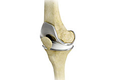
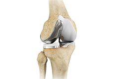
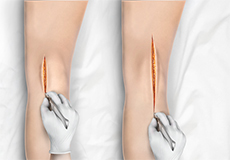
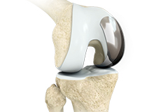
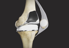
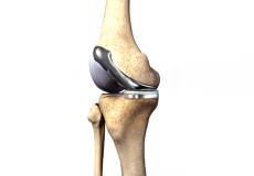
Total Knee Replacement
Total knee replacement, also called total knee arthroplasty, is a surgical procedure in which the worn out or damaged surfaces of the knee joint are removed and replaced with an artificial prosthesis.
Unicompartmental/Partial Knee Replacement
Unicompartmental knee replacement is a minimally invasive surgery in which only the damaged compartment of the knee is replaced with an implant. It is also called a partial knee replacement.
Minimally Invasive Knee Joint Replacement
Total knee replacement is a very successful surgical treatment for knee arthritis. Over the years, minimally invasive knee replacement surgical techniques have been developed to lessen tissue trauma and improve patient outcomes.
Patient Specific Knee Replacement
Patient Specific Knee Replacement is a newer technology in total knee replacement surgery. It is an advanced procedure using an individualized patient-specific knee implant for replacement of all three components of the knee. The difference with patient specific knee replacement from other knee replacement surgeries is the use of an MRI scan prior to the surgery that provides a clear view of the shape and structure of the different components of the joint.
Outpatient Unicondylar Knee Replacement
A unicondylar knee replacement, also known as unicompartmental or partial knee replacement, is a procedure to replace a portion of the damaged knee joint with a prosthetic implant to relieve pain and improve function of the knee joint.
Tricompartmental Knee Replacement
Tricompartmental knee replacement, also called total knee arthroplasty, is a surgical procedure in which the worn-out or damaged surfaces of the knee joint are removed and replaced with artificial parts.
Custom Knee Replacement
Custom Knee Replacement is an advanced surgical procedure in which the damaged knee joint is replaced by a customized implant, specifically designed to match the unique size and shape of each patient’s knee.
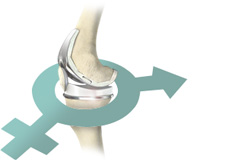
Gender-Specific Knee Replacement
Gender-specific knee replacement, also known as woman’s knee replacement, is a surgical procedure in which gender-specific knee implants specifically designed for a male or female are used for total knee replacement surgery.